Histology - Epoxy Resin TAAB PREMIX
Embryos set in epoxy resin were serially sectioned at 1μ thickness.
Protocol
Embryo collection and preparation
The embryo was prepared for sectioning as described in Embryo collection - Epoxy resin
Sectioning
The embryo for sectioning was cut out with a small hacksaw, trimmed with a single edge razor blade and clamped in a vice type ultratome chuck. The chuck was fixed to the ultratome and the block face trimmed with a glass knife until it was almost at the embryo.
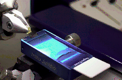
The glass knife was then replaced with a Diatome Histo Jumbo Diamond knife (M.J.F. Blumer, P Gahleitner, T Natzt, C. Handl, B Ruthensteiner : Strips of semi-thin sections: an advanced method with a new type of diamond knife. Journal of Neuroscience Methods 120, pp.11-16, 2002.
NB Bostik/Evo-stik impact adhesive diluted 1:1 in toluene was used instead of Pattek adhesive.
Serial 2μ sections were cut onto distilled water in the trough. Contact adhesive was used on the top and bottom edge of the block to allow ribbons of sections to be cut. Ribbons of sections were transferred to a pool of distilled water on a cleaned glass slide (2-3 per slide). The slide was placed on a hot plate at 70oC and the sections allowed to stretch out fully for 2 mins. The ribbons of sections were orientated on the slide and the water withdrawn with a fine tipped pastette.
The slide was dried vertically overnight at room temperature then heated on a hot plate at 70oC for 1 hr to adhere the sections to the slide prior to staining.
| Floating out |
 |
Staining
The slides were stained with 1% Toluidine Blue in 1% Borax diluted 1:20 in distilled water in a coplin jar in a water bath at 60oC for 30 seconds. The slides were then rinsed in distilled water and allowed to air dry before mounting them in microscope oil with a coverslip.
Image Capture
After staining the slides were available for image capture


