Movies Protocols
The OPT camera Retiga Ex (monochrome) produces a movie of 400 images (tif) to show a 2D rotational movie. Subsequent scans can be superimposed and falsely coloured for differentiation purposes.
Surface water movies
As an alternative to using BABB as a clearing agent we have developed a method to scan samples in water which allows the viewer to see the surface of the embryo.
We have also found these outputs to be useful in helping to Theiler stage embryos.
We have morphed these outputs together using Xmorph to show the development of mouse embryos from Ts17 to Ts28.
We have also used Xmorph on staged sections Ts17 - Ts28 from developing kidneys giving an insight as to how the kidney develops through these stages.
The example surface water movies can be viewed in the gallery.
Methodology
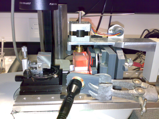
Embryos are fixed in Bouins fixative and briefly washed before setting the embryo in 1% low melting point agarose. As the embryo is to be scanned in water it is vital not to have any flat sides visible to the camera so a custom made circular device is used to produce a perfectly round mount. The block is stuck to a metal mount and is scanned in water with light shone onto the object.
 |
| embryo scanning |
For falsely coloured scans the embryos are fixed in 4% PFA, the embryo is scanned 3 times using a blue, red, green colour additive filter sets (Andover Corporation) . The 3 scans are superimposed in IPlabs to produce a movie. See gallery.
Below a comparison of embryos fixed in PFA and Bouins.
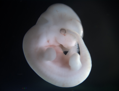
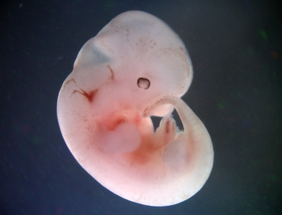
Bouins fixative- showing the surface of the embryo and an embryo fixed in PFA which gives an indication of vasculature. * link to movie*
 |
 |
| Bouins | PFA |
The embryo photographs featured on our EMA stage selector from TS15 onwards are from a screengrab either from monochrome or falsely coloured water scan.


